Colloid Cyst
A colloid cyst is a type of brain cyst. A brain cyst is an abnormal fluid-filled sac in the brain. The cyst contains a thick gel-like substance called colloid. A colloid cyst is generally benign (not cancer). Benign also means that the growth doesn't spread to other parts of the body. Often, a brain cyst starts before birth. A colloid cyst may be present through childhood but not large enough to cause symptoms until adulthood.
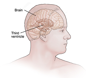
How a colloid cyst can cause problems
A colloid cyst often forms near the third ventricle of the brain. This is a space in the center of the brain that holds cerebrospinal fluid (CSF). CSF is a clear fluid that protects and cushions your brain and spine. A cyst can block the flow of CSF. It can also cause positional headaches. These are headaches that occur when you are in a certain position. Untreated pressure in the brain caused by colloid cysts can cause bleeding in the brain or sudden death.
What causes a colloid cyst?
Researchers are not sure what causes a colloid cyst. The cyst may start to form when a baby’s central nervous system is developing in the womb.
Symptoms of a colloid cyst
A colloid cyst may not cause symptoms unless it grows large, usually after age 30. The symptoms depend on which part of the brain the cyst is growing in. Symptoms can occur a bit differently in each person but can include:
A colloid cyst may cause CSF to build up on the brain. This is called hydrocephalus. It can cause symptoms such as:
-
Confusion.
-
Changes in personality.
-
Double or blurred vision.
-
Sleepiness.
-
Balance and coordination problems.
-
Nausea or vomiting.
-
Loss of bladder control.
-
Trouble walking or talking.
Diagnosing a colloid cyst
A colloid cyst may be found when it shows up on an imaging scan done for another reason. In other cases, you may have symptoms from the cyst. Your health care provider may refer you to a neurologist. This is a specialized provider who diagnoses and treats diseases of the central nervous system. You may also be referred to a neurosurgeon who does brain or spinal cord surgery.
Your health care provider will ask about your medical history and symptoms. They will give you a physical and neurologic exam. Imaging tests may be done to look at the brain. Contrast material may be used to help show more detail in the images. The tests may include:
-
CT scan. This is a test that uses a series of X-rays and a computer to create detailed images of the inside of the body. Scans may be done of your brain and spinal cord.
-
MRI. This test uses large magnets and a computer to create detailed images of the body without the use of X-rays. MRI scans of your brain and spinal cord may be done to get more information about the cyst and nearby tissues.
Scans may be repeated over time to see if the cyst is growing.
Treatment for a colloid cyst
A colloid cyst may stay the same size for decades or may grow bigger over time. Cysts that block CSF flow can be life-threatening if untreated. A colloid cyst can be emptied or removed with surgery. The size and location of the cyst, as well as any symptoms, will determine if surgery is needed.
Hydrocephalus caused by a colloid cyst is treated with a shunt. A shunt is a small drainage tube put in the brain, which then drains extra CSF from the brain ventricles into the abdomen. This helps to remove fluid and relieve pressure.
When to call your doctor
Contact your doctor or seek medical care right away if you have: