Understanding Aortic Valve Regurgitation
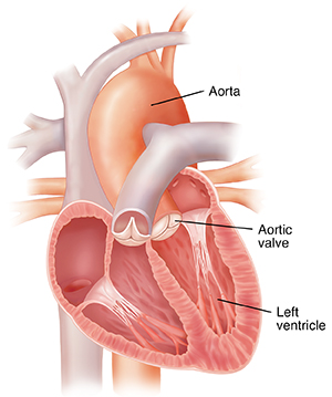
Aortic valve regurgitation is when the aortic valve leaks. The aortic valve is one of the heart’s four valves. It's on the left side of the heart. It sits between the left lower chamber (left ventricle) and the large blood vessel that sends oxygenated blood to the body (aorta).

What is aortic valve regurgitation?
Valves in the heart help blood flow through the heart in the right direction. The aortic valve keeps blood flowing from the left ventricle to the aorta. When the heart squeezes, the valve is pushed open to let blood flow out of the heart. When the heart relaxes, the valve closes and prevents blood from flowing backward into the heart.
With aortic valve regurgitation, the aortic valve does not work right. Some blood leaks back into the left ventricle when the valve closes. As a result, less blood flows forward into the aorta to go to the body. The backflow adds to the volume in the heart (volume overload). This can quickly lead to high pressures in the heart and lungs, resulting in shortness of breath. Over time, the heart works harder to pump blood, which can lead to heart failure. This may occur with moderate to severe aortic valve regurgitation.
There are two types of aortic valve regurgitation:
-
Acute aortic valve regurgitation. The valve suddenly starts to leak. The heart doesn’t have time to get used to the leaking. This is a medical emergency. Symptoms often start suddenly and are severe.
-
Chronic aortic valve regurgitation. The valve becomes leaky over time. The heart has time to get used to the leak. Symptoms may start gradually and get worse over time because the heart and the body are able to adjust to the change in blood flow.
What causes aortic valve regurgitation?
The causes of aortic valve regurgitation can include:
-
Weakening of the valve from aging.
-
High blood pressure.
-
Valve defects at birth (congenital).
-
Infections, such as an infection of the heart lining (infective endocarditis).
-
Valve damage from untreated strep infections, such as strep throat (rheumatic heart disease).
-
Problems with the aorta, such as a tear in the vessel wall (dissection) or weakening and ballooning out of the vessel wall (aneurysms).
-
Injuries to the chest.
Symptoms of aortic valve regurgitation
You may not have any symptoms if you have mild aortic regurgitation. If your condition is more severe or if it gets worse over time, you may have symptoms such as:
-
Shortness of breath with exercise, activity, or exertion, or when lying down.
-
Chest pain.
-
Tiredness (fatigue).
-
Feeling the heart beat faster or harder (palpitations).
-
Swelling in your legs, belly (abdomen), and the veins in your neck.
Sudden severe aortic valve regurgitation is a medical emergency. Call 911 right away. Signs and symptoms may include:
-
Pale skin, loss of consciousness, fast breathing, and fast heartbeat (symptoms of shock).
-
Severe dizziness.
-
Severe shortness of breath.
-
Chest pain.
-
Abnormal heart rhythm (arrhythmia).
Diagnosing aortic valve regurgitation
Your health care provider will ask you about your medical history and symptoms. They will examine you. The exam will include listening to your heart with a stethoscope. This will let your health care provider hear if you have an abnormal heart sound (heart murmur). You may also have tests, such as:
-
An electrocardiography (ECG). Sticky pads (electrodes) are put on your chest. Wires are attached and connected to a machine. The machine checks your heart rhythm.
-
A chest X-ray. This test uses a small amount of radiation to make pictures of your heart and lungs. This is done to check for any changes such as an enlarged heart or fluid in the lungs.
-
A transthoracic echocardiography (TTE). Sound waves are used to make pictures of your heart using a wand (transducer) placed over your chest.
-
A transesophageal echocardiography (TEE). This is another way of using sound waves to make pictures of your heart. The health care provider passes a transducer down your esophagus to get pictures. This type of test may be used when certain pictures of the heart are needed. For example, it may be used to check for problems with the aorta.
-
A CT scan. This test uses a series of X-rays and a computer to create detailed pictures of your heart and aorta.
-
A cardiac MRI (CMR). Magnets, radio waves, and a computer are used to make detailed pictures of your heart and aorta. It does not use X-rays.
-
An exercise stress test. During this test, an ECG is done while you exercise. This checks how your heart reacts to stress. Pictures of the heart may be taken during this test.
-
A cardiac catheterization (heart cath), an aortography, or a coronary angiography. These are invasive tests, or tests in which a doctor puts something in your skin or body. They may be needed if other test results are not clear. They may also be done if surgery is planned.